
Fitxer:Fundus of patient with retinitis pigmentosa, mid stage.jpg
Mida d'aquesta previsualització: 699 × 599 píxels. Altres resolucions: 280 × 240 píxels | 560 × 480 píxels | 871 × 747 píxels.
Fitxer original (871 × 747 píxels, mida del fitxer: 107 Ko, tipus MIME: image/jpeg)
| Aquest fitxer i la informació mostrada a continuació provenen del dipòsit multimèdia lliure Wikimedia Commons. |
| Descripció |
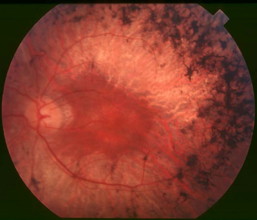
English: Figure 2. Fundus of patient with retinitis pigmentosa, mid stage (Bone spicule-shaped pigment deposits are present in the mid periphery along with retinal atrophy, while the macula is preserved although with a peripheral ring of depigmentation. Retinal vessels are attenuated.) Hamel Orphanet Journal of Rare Diseases 2006 1:40 doi:10.1186/1750-1172-1-40 |
| Data | |
| Font | Retinitis pigmentosa by Christian Hamel |
| Autor | Christian Hamel |
| Permís (Com reutilitzar aquest fitxer) |
© 2006 Hamel; licensee BioMed Central Ltd. This is an Open Access article distributed under the terms of the Creative Commons Attribution License (https://creativecommons.org/licenses/by/2.0), which permits unrestricted use, distribution, and reproduction in any medium, provided the original work is properly cited. |
Aquest fitxer està disponible sota la llicència Creative Commons Reconeixement 2.0 Genèrica.
- Sou lliure de:
- compartir – copiar, distribuir i comunicar públicament l'obra
- adaptar – fer-ne obres derivades
- Amb les condicions següents:
- reconeixement – Heu de donar la informació adequada sobre l'autor, proporcionar un enllaç a la llicència i indicar si s'han realitzat canvis. Podeu fer-ho amb qualsevol mitjà raonable, però de cap manera no suggereixi que l'autor us dóna suport o aprova l'ús que en feu.
Historial del fitxer
Cliqueu una data/hora per veure el fitxer tal com era aleshores.
| Data/hora | Miniatura | Dimensions | Usuari/a | Comentari | |
|---|---|---|---|---|---|
| actual | 12:17, 2 des 2017 | | 871 × 747 (107 Ko) | Doc James | Cropped 27 % horizontally and 7 % vertically using CropTool with precise mode. |
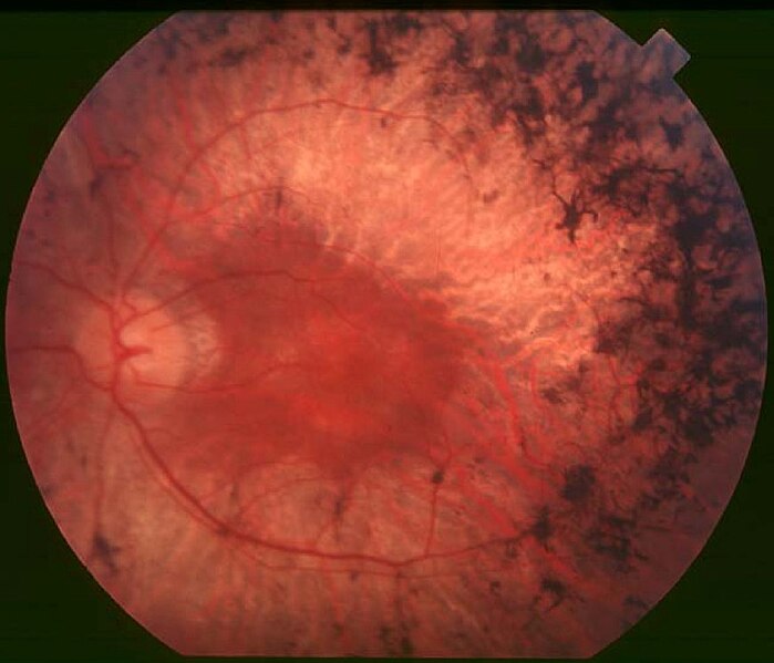
| 15:52, 22 set 2009 |  | 1.200 × 799 (126 Ko) | CopperKettle | {{Information |Description={{en|1=Figure 2. Fundus of patient with retinitis pigmentosa, mid stage (Bone spicule-shaped pigment deposits are present in the mid periphery along with retinal atrophy, while the macula is preserved although with a peripheral |
Ús del fitxer
La pàgina següent utilitza aquest fitxer:
Ús global del fitxer
Utilització d'aquest fitxer en altres wikis:
- Utilització a ar.wikipedia.org
- Utilització a bs.wikipedia.org
- Utilització a da.wikipedia.org
- Utilització a en.wikipedia.org
- Utilització a en.wikiversity.org
- Utilització a es.wikipedia.org
- Utilització a eu.wikipedia.org
- Utilització a fa.wikipedia.org
- Utilització a fi.wikipedia.org
- Utilització a fr.wikipedia.org
- Utilització a he.wikipedia.org
- Utilització a hy.wikipedia.org
- Utilització a it.wikipedia.org
- Utilització a ko.wikipedia.org
- Utilització a la.wikipedia.org
- Utilització a or.wikipedia.org
- Utilització a outreach.wikimedia.org
- Utilització a pl.wikipedia.org
- Utilització a pt.wikipedia.org
- Utilització a ru.wikipedia.org
- Utilització a sl.wikipedia.org
- Utilització a sr.wikipedia.org
- Utilització a sv.wikipedia.org
- Utilització a th.wikipedia.org
- Utilització a tr.wikipedia.org
- Utilització a tt.wikipedia.org
- Utilització a uk.wikipedia.org
- Utilització a vi.wikipedia.org
Vegeu més usos globals d'aquest fitxer.