
Fitxer:GFP Superresolution Christoph Cremer.JPG
GFP_Superresolution_Christoph_Cremer.JPG (538 × 389 píxels, mida del fitxer: 156 Ko, tipus MIME: image/jpeg)
| Aquest fitxer i la informació mostrada a continuació provenen del dipòsit multimèdia lliure Wikimedia Commons. |
Resum
| Descripció |
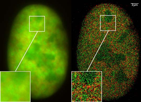
GFP superresolution, optical nanoscopy ( Christoph Cremer, emeritus at Heidelberg university [1]) View of a nucleus of a bone cancer cell: using normal high resolution fluorescence microscopy, it is not possible to distinguish details of its structure (image on the left). Using the two Color Localization Microscopy 2CLM (image on the right) it is possible to localize 70,000 histone molecules (red: RFP-H2A) and 50,000 chromatin remodeling proteins (green: GPF-Snf2H) in a field of view of 470 µm2 with an optical depth of 600 nm. Common fluorescence markers were used. 2CLM is the only optical nanoscopy method that allows position based co-localization of single molecules at high density in a wide field of view using conventional fluorescent proteins such as GFP, YFP, RFP, or other conventional fluorochromes. Due to its high optical single molecule resolution, 2CLM allows significantly more precise analyses of potential protein interactions than FRET-(Fluorescence Resonance Energy Transfer) technology, which is at present the preferred method for such investigations. This is of particular significance in studies of biomolecular machines (BMMs) within cells: Single BMMS can be analysed, including the number of molecules of a given type; distances between proteins in these BMMs often are substantially greater than those that can be analyzed by FRET (restricted to a maximum distance of only a few nm). Possible to use conventional, well established and inexpensive fluorescent dyes, from the GFP group, and its dye variants, to the well-known Alexa and fluorescein dyes. Fundamental to SPDMphymod are blinking phenomena (flashes of fluorescence), induced by reversible bleaches (metastable dark states). Individual molecules of the same spectral emission color can be detected. Publikation: Manuel Gunkel, Fabian Erdel, Karsten Rippe, Paul Lemmer, Rainer Kaufmann, Christoph Hörmann, Roman Amberger and Christoph Cremer: Dual color localization microscopy of cellular nanostructures. In: Biotechnology Journal, 2009, 4, 927-938. ISSN 1860-6768 |
| Data | 073009 |
| Font | Treball propi |
| Autor | Andy Nestl |
| Permís (Com reutilitzar aquest fitxer) |
Gallery
- Super Resolution Microscopy - Localisation Microscopy
-
 Breast Cancer Cells: 3D Dual Color Super Resolution Microscopy of Her2 and Her3 & cluster calculations
 -
 Single YFP molecule detection in a human cancer cell. Typical distance measurements 15 nm
 -
 Co- localisation microscopy with GFP and RFP fusion proteins (nucleus of a bone cancer cell) 120.000 localized molecules in a widefield area(470 µm2)
 -
 Label-free Localisation Microscopy SPDM - Super Resolution Microscopy reveals prior undetebable intracellular structures
 -
 Investigation of human eye tissue, affected by macular degeneration AMD
 -
 Virus Super Resolution Microscopy SPDM Cremer/Wege labs


Llicència
- Sou lliure de:
- compartir – copiar, distribuir i comunicar públicament l'obra
- adaptar – fer-ne obres derivades
- Amb les condicions següents:
- reconeixement – Heu de donar la informació adequada sobre l'autor, proporcionar un enllaç a la llicència i indicar si s'han realitzat canvis. Podeu fer-ho amb qualsevol mitjà raonable, però de cap manera no suggereixi que l'autor us dóna suport o aprova l'ús que en feu.
- compartir igual – Si modifiqueu, transformeu, o generareu amb el material, haureu de distribuir les vostres contribucions sota una llicència similar o una de compatible com l'original

|
S'autoritza la còpia, la distribució i la modificació d'aquest document sota els termes de la llicència de documentació lliure GNU versió 1.2 o qualsevol altra versió posterior que publiqui la Free Software Foundation; sense seccions invariants, ni textos de portada, ni textos de contraportada. S'inclou una còpia d'aquesta llicència en la secció titulada GNU Free Documentation License. |
Resum
- ↑ https://www.physik.uni-heidelberg.de/personen/lsf.php?details=1537 |titel=Fakultät für Physik und Astronomie |abruf=2020-10-01
Historial del fitxer
Cliqueu una data/hora per veure el fitxer tal com era aleshores.
| Data/hora | Miniatura | Dimensions | Usuari/a | Comentari | |
|---|---|---|---|---|---|
| actual | 14:14, 30 jul 2009 | | 538 × 389 (156 Ko) | Andy Nestl | {{Information |Description=GFP superresolution, optical nanoscopy (Christoph Cremer) |Source=Own work by uploader |Date=073009 |Author=Andy Nestl |Permission=given by Christoph Cremer, University of Heidelberg |other_versions= }} |
Ús del fitxer
La pàgina següent utilitza aquest fitxer:
Ús global del fitxer
Utilització d'aquest fitxer en altres wikis:
- Utilització a ar.wikipedia.org
- Utilització a be.wikipedia.org
- Utilització a bn.wikipedia.org
- Utilització a cs.wikipedia.org
- Utilització a de.wikipedia.org
- Utilització a en.wikipedia.org
- Utilització a en.wikibooks.org
- Utilització a eo.wikipedia.org
- Utilització a fa.wikipedia.org
- Utilització a fr.wikipedia.org
- Utilització a gl.wikipedia.org
- Utilització a he.wikipedia.org
- Utilització a it.wikipedia.org
- Utilització a mai.wikipedia.org
- Utilització a ne.wikipedia.org
- Utilització a nl.wikipedia.org
- Utilització a pl.wikipedia.org
- Utilització a pt.wikipedia.org
- Utilització a sv.wikipedia.org
- Utilització a ta.wikipedia.org
- Utilització a vi.wikipedia.org
- Utilització a zh.wikipedia.org